


Advanced 3D dental imaging techniques offer detailed, three-dimensional visualizations of intricate mouth structures, surpassing traditional 2D X-rays. This technology aids in accurately detecting subtle abnormalities in TMJs and sinus cavities, benefiting family and restorative dentistry practices through early identification of disorders. In pediatric dentistry, it promotes less invasive diagnosis for optimal oral health development, while also enhancing treatment planning for procedures like tooth extractions and clear aligner treatments.
“Unveiling the intricacies of oral health with 3D dental imaging, this article explores its revolutionary role in TMJ (Temporal Mandibular Joint) and sinus evaluations. By delving into the advanced technology, we uncover how it provides unprecedented detail for precise diagnoses. Understanding TMJ and sinus conditions is paramount, and 3D imaging offers a game-changer in visualization. This comprehensive guide details the advantages, from improved accuracy to reduced scan times, and walks you through the entire process, ensuring a clear understanding of what to expect.”
- Understanding TMJ and Sinus Conditions
- The Advantages of 3D Dental Imaging
- The Process and What to Expect
Understanding TMJ and Sinus Conditions

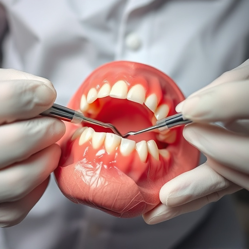
TMJ (Temporomandibular Joint) and sinus conditions are complex oral health issues that often require advanced diagnostic techniques to accurately assess and treat. 3D dental imaging has emerged as a game-changer in this field, offering clear, detailed visualizations of the mouth’s intricate structures, including the TMJ and sinus cavities. This innovative technology goes beyond traditional 2D X-rays, providing dentists with a more comprehensive understanding of these conditions.
By capturing precise, three-dimensional data, 3D dental imaging allows for the detection of subtle abnormalities that may be obscured on standard radiographs. This is especially beneficial in family dentistry and restorative dentistry practices, where early identification of TMJ disorders or sinus issues can lead to more effective treatment planning. For children’s dentistry patients, this advanced technology ensures a less invasive approach to diagnosis, promoting optimal oral health development.
The Advantages of 3D Dental Imaging
3D dental imaging offers a transformative approach to oral health evaluations, providing a detailed and comprehensive view of the complex structures within the mouth. This advanced technology surpasses traditional 2D radiography by delivering highly accurate, three-dimensional models of teeth, jaws, and surrounding tissues. Dentists can now detect even subtle abnormalities that might be missed on conventional images.
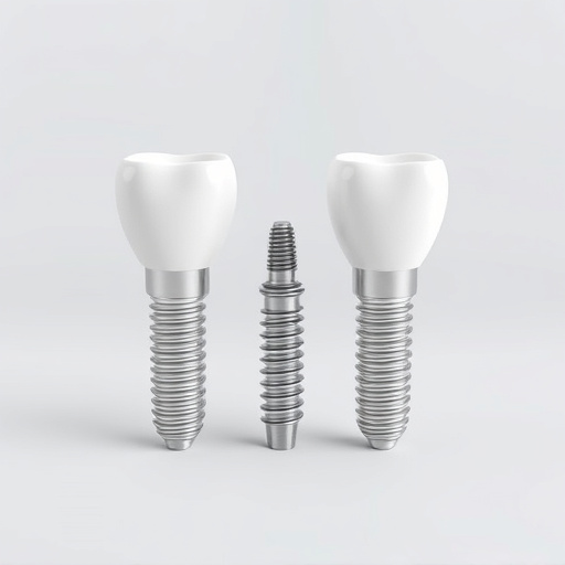
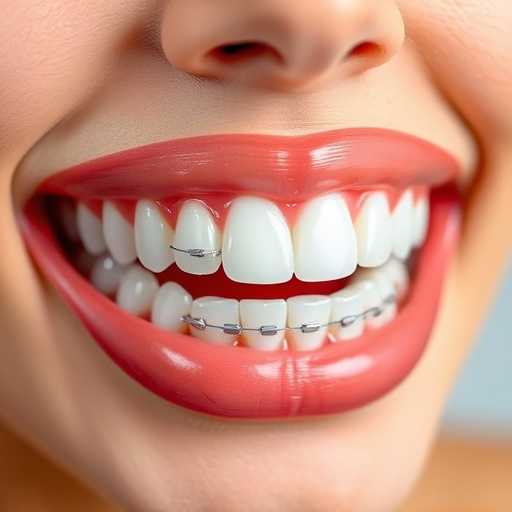
One of the significant advantages is its ability to facilitate precise planning for tooth extractions and clear aligner treatments in family dentistry. By visualizing the entire oral cavity in 3D, dental professionals can make informed decisions, ensuring better outcomes and reduced treatment time. This technology revolutionizes the way dental care is delivered, offering a clearer path to effective and efficient patient management.
The Process and What to Expect
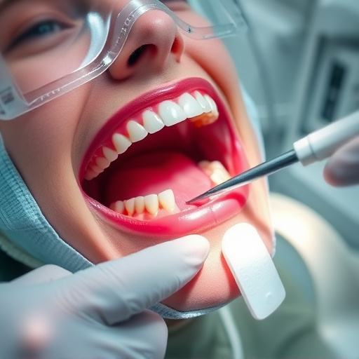
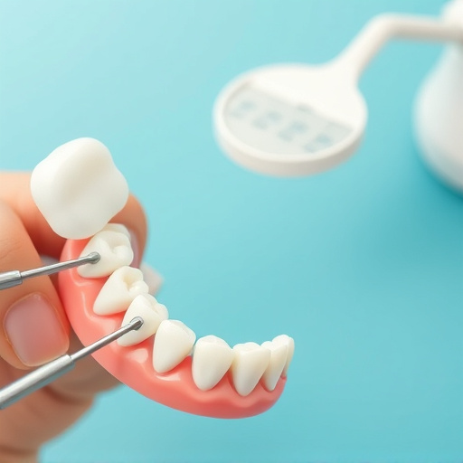
The process of 3D dental imaging for TMJ (Temporal Mandibular Joint) and sinus evaluations is both advanced and non-invasive, offering a detailed glimpse into your oral health. It begins with a scan using specialized equipment that captures multiple images from various angles, creating a precise, three-dimensional model of your jaw joint and surrounding areas, including sinuses. This technology, often employed in family dentistry or general dentistry practices, allows dentists to diagnose conditions like TMJ disorders, sinus infections, or even plan for dental crowns with remarkable accuracy.
During the scan, you’ll be asked to sit or stand still while the imaging device rotates around your head, taking rapid successive images. The entire process is comfortable and usually takes just a few minutes. Afterwards, these images are processed using specialized software that reconstructs them into a detailed 3D model, providing dentists with a clear view of intricate structures not visible through traditional 2D imaging. This advanced technique ensures patients can receive personalized treatment plans, enhancing the precision and effectiveness of procedures, including those involving dental crowns.
3D dental imaging has emerged as a game-changer in TMJ and sinus evaluations, offering unprecedented precision and detailed insights. By providing a comprehensive view of oral structures, this advanced technology enables dentists to diagnose conditions more effectively. The advantages are clear: faster, more accurate assessments lead to improved treatment planning and outcomes. Understanding these benefits and the simple process involved paves the way for better patient care and enhanced dental practices, making 3D dental imaging an indispensable tool in modern dentistry.